
01 · Seed

NanoAMi · Evidence by component
What supports each part of NanoAMi?
The biological argument is tissue-informed. Here the fibroblast-rich Seed, collagen Soil and platelet-rich plasma Signal are each shown with their published evidence before the combined protocol is described.
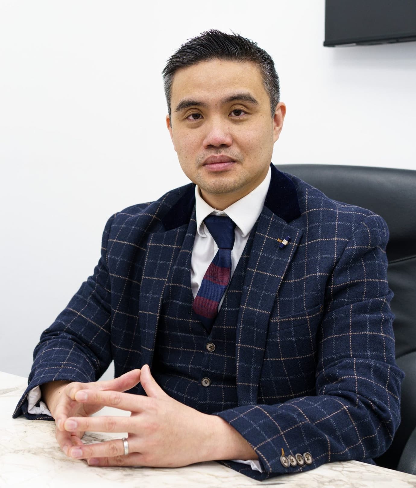 Reviewed byProfessor Paul Lee MBBch, FRCS (Tr & Orth), PhDCartilage and joint preservation expertise
Quick answer
NanoAMi uses autologous fibroblast-rich micrografts, an injectable type-I collagen scaffold and platelet-rich plasma, delivered by injection without an operation. Meniscus cell biology, human collagen meniscus implant studies and a randomised trial of PRP-augmented meniscal repair support each part. Outcomes for the complete technique are recorded at the clinic.
Verified across three networks
Trusted by patients on Doctify, Google and Top Doctors
A transparent evidence map
Three components. Three evidence questions.
Each part is assessed against the meniscus target. Evidence for one component is never presented as proof of the complete protocol.
02 · Soil
Type-I collagen scaffold
03 · Signal
Platelet-rich plasma
01 · Seed
Autologous fibroblast-rich micrografts
A small hair-follicle-bearing tissue sample is mechanically processed on the day into an autologous micrograft suspension containing fibroblast-rich dermal tissue and native matrix.
How directly does the evidence apply?
Hair-follicle niches contain dermal precursor populations with neural-crest-like properties and multi-lineage potential under laboratory conditions. Human meniscus contains fibroblast-like and fibrochondrocyte populations. These findings are the biological basis for a fibroblast-rich Seed in meniscus tissue.
Target-tissue evidenceHuman cell isolation and in-vitro collagen-scaffold study.
Human meniscus cells include fibroblast-like populations
Nakata K et al. Clinical Orthopaedics and Related Research. 2001;(391 Suppl):S208–S218.
Cells isolated from human meniscus included elongated fibroblast-like cells and attached to a porous collagen scaffold while expressing meniscal matrix genes.
Why it belongs here: Supports the fibroblast and collagen biology of the target tissue, but does not test hair-follicle micrografts or a percutaneous NanoAMi treatment.
Read the published sourceMechanistic evidenceLineage-tracing and laboratory study.
Hair-follicle dermal precursors from distinct developmental origins
Jinno H et al. Stem Cells. 2010;28(11):2027–2040.
Follicle-associated dermal precursors from face and trunk had different developmental origins but similar neural-crest-like properties and multi-lineage behaviour.
Why it belongs here: Supports the choice of the donor niche and corrects the over-broad claim that every hair-follicle cell is neural-crest-derived. It is not meniscus treatment evidence.
Read the published source02 · Soil
Injectable type-I collagen scaffold
A three-dimensional type-I collagen framework intended to retain the delivered biology at the selected meniscus target.
How directly does the evidence apply?
Human meniscus cells have been studied on type-I collagen scaffolds, and a different meniscus-specific collagen implant has clinical trial evidence. Professor Lee selects ChondroFiller as the injectable scaffold; its published product evidence is in articular cartilage, and the meniscus implant trials support the scaffold principle in the target tissue.
Mechanistic evidenceHuman in-vitro cell and scaffold study.
Human meniscus cells on type-I collagen scaffolds
Adesida AB et al. PLoS ONE. 2012;7(6):e39365.
Human meniscal fibrochondrocytes formed matrix after culture on a collagen scaffold, with phenotype affected by culture conditions.
Why it belongs here: Target-tissue support for a collagen scaffold concept; not a study of the chosen injectable product or of NanoAMi.
Read the published sourceIndirect evidenceRandomised controlled trial involving 311 patients.
Collagen meniscus implant compared with partial meniscectomy
Rodkey WG et al. Journal of Bone and Joint Surgery Am. 2008;90(7):1413–1426.
A meniscus-specific collagen implant was assessed as a surgical scaffold after loss of meniscal tissue.
Why it belongs here: Supports the broader meniscus-scaffold principle only. The implant, surgery and indication are not ChondroFiller or NanoAMi.
Read the published source03 · Signal
Autologous platelet-rich plasma
A platelet concentrate prepared from the patient’s blood that adds platelet-derived growth factors to the combined pathway.
How directly does the evidence apply?
PRP has a randomised placebo-controlled trial and cohort studies as an augment to meniscal repair, plus small-series experience as a non-operative injection. Those studies test PRP without the Seed or Soil; in NanoAMi it is the Signal delivered alongside both.
Target-tissue evidenceProspective double-blind randomised placebo-controlled trial, 37 patients, bucket-handle tears.
PRP-augmented meniscal repair: randomised placebo-controlled trial
Kaminski R et al. BioMed Research International. 2018;2018:9315815.
Repairs augmented with PRP healed more often than repairs without it, with fewer failures at follow-up.
Why it belongs here: Human target-tissue evidence that the patient’s own platelets support meniscal healing. It studied PRP around surgical repair, not the non-operative NanoAMi combination.
Read the published sourceTarget-tissue evidenceComparative cohort study of 550 meniscal repairs with a minimum two-year follow-up.
PRP reduces failure risk for isolated meniscal repairs
Everhart JS et al. American Journal of Sports Medicine. 2019;47(8):1789–1796.
For isolated meniscal repairs, PRP augmentation lowered the repair failure rate compared with repairs without it.
Why it belongs here: Human target-tissue evidence that the patient’s own platelets support meniscal healing after repair; PRP around surgery, not the non-operative combination.
Read the published sourceTarget-tissue evidenceComparative clinical study of arthroscopic repair with and without PRP.
PRP in arthroscopic repair of discoid lateral meniscus tears
Dai WL et al. BMC Musculoskeletal Disorders. 2019;20:113.
Repairs augmented with PRP had better functional scores and healing on follow-up imaging than repairs without it.
Why it belongs here: Further human evidence that PRP augmentation supports meniscal healing in the target tissue.
Read the published sourceIndirect evidenceTen-patient case series; no control group.
Percutaneous PRP for degenerative meniscus lesions
Blanke F et al. Muscles, Ligaments and Tendons Journal. 2015;5(3):162–166.
Six patients reported improved pain and activity; four later proceeded to surgery.
Why it belongs here: Relevant to non-operative meniscus injection with PRP alone; it does not include the Seed or Soil components.
Read the published sourceThe combined protocol
Three established principles, one technique
NanoAMi is Professor Lee’s clinician-developed technique for combining fibroblast-rich micrografts, collagen support and an autologous signal. In the standard pathway described on this site it is delivered percutaneously without an operation, arthroscopy or stitches. Outcomes for the complete technique are recorded at the clinic.
Each component is used within its own evidence base. Professor Lee brings them together in one image-guided session, and outcomes for the complete technique are recorded at the clinic so that it is judged on its own results as well as on the evidence for its parts.

Questions about the NanoAMi evidence
What evidence supports NanoAMi?
Meniscus contains fibroblast-like cells, which the fibroblast-rich Seed is chosen to match; collagen meniscus implants have human clinical evidence for the scaffold principle; and PRP has a randomised placebo-controlled trial as an augment to meniscal repair. Each study is shown with how directly it applies, and outcomes for the complete technique are recorded at the clinic.
What supports the fibroblast-rich Seed?
Hair-follicle dermal precursors show neural-crest-like and multi-lineage properties in laboratory research, and meniscus contains fibroblast-like cell populations.
Why use a collagen scaffold in the meniscus?
Human evidence for collagen meniscus implants supports the scaffold principle in the meniscus. ChondroFiller’s published clinical evidence is in articular cartilage, and it provides the injectable type-I collagen framework used as the Soil.
Can NanoAMi replace meniscus stitches?
No. It may be considered for selected stable, viable damage where mechanical fixation is not required. A displaced tear, root tear or unstable repairable tear remains a structural problem and may require surgery.
Still have more specific concerns?
Free Discovery CallOptions that our doctors may discuss include NanoACi, ChondroFiller, Mytocel MSK, joint replacement, established conservative care or surgery, depending on examination and imaging.
Discuss the evidence, limitations and alternatives
Professor Lee can explain how the evidence applies to your meniscus, where the rationale is extrapolated and whether this pathway is proportionate.